Next: Brain scans to identify children at high risk of developing multiple sclerosis (MS) before symptoms appear
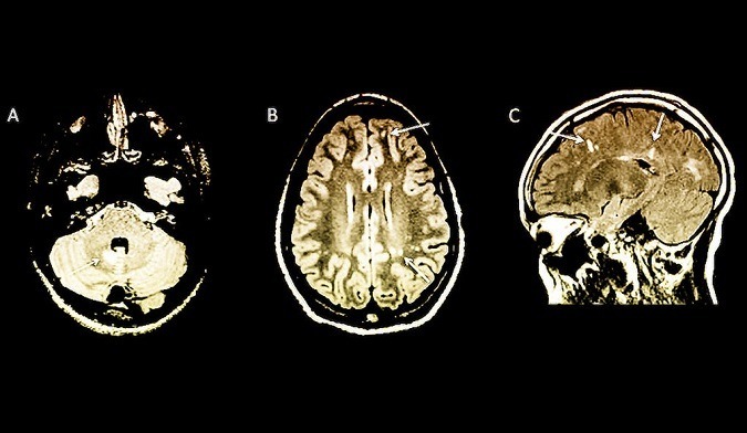
White arrows indicate abnormalities on MRI brain scans in children with no symptoms of multiple sclerosis.
______________________________
MS risk in children spotted with MRI brain scans (Yale News):
“By the time multiple sclerosis (MS) is diagnosed in children, it may be difficult to prevent the disabilities and relapses that come with the disease. In a new Yale School of Medicine study, researchers examined MRI brain scans to identify children at high risk of developing MS before symptoms appear, which may lead to earlier diagnosis and treatment…the study of 38 children at 16 sites in six countries showed that the MRIs can reveal changes in the brain associated with MS before the clinical symptoms of the disease appear in children.
The children in the study all underwent MRI scans for other reasons, most commonly headache, but the MRIs unexpectedly revealed signs of MS. Having MRI findings of MS without any symptoms of the disease has been termed radiologically isolated syndrome (RIS) and previously had only been seen in adults.
“For the first time we have proposed a definition of RIS in children,” said lead author Naila Makhani, M.D., assistant professor of pediatrics and neurology at Yale School of Medicine. “Children with RIS may represent a high-risk group of children that needs to be followed more closely for the later development of clinical multiple sclerosis.”
The Study
Radiologically isolated syndrome in children (Neurology: Neuroimmunology & Neuroinflammation). From the abstract:
- Objective: To describe clinical and radiologic outcomes of children with incidental findings on neuroimaging suggestive of CNS demyelination (termed “radiologically isolated syndrome” or RIS).
- Methods: Clinical and radiologic data were obtained from a historical cohort of children with no symptoms of demyelinating disease who had MRI scans that met the 2010 MRI criteria for dissemination in space for MS.
- Results: We identified 38 children (27 girls and 11 boys) with RIS now being prospectively followed at 16 sites in 6 countries. The mean follow-up time was 4.8 ± 5.3 years. The most common reason for initial neuroimaging was headache.
- Conclusions: We describe the clinical characteristics and outcomes of children with incidental MRI findings highly suggestive of CNS demyelination. Children with RIS had a substantial risk of subsequent clinical symptoms and/or radiologic evolution. The presence of oligoclonal bands in CSF and spinal cord lesions on MRI were associated with an increased risk of a first clinical event.
News in Context
- Next: Harnessing brain scans to personalize autism-related behavioral interventions
- Can brain scans identify ADHD and help predict treatment response?
- Brain waves help predict stress-related sleep problems
- Cognitive therapy or medication? Brain scans may help personalize treatments
- Study: Transcranial Direct Current Stimulation (tDCS) can reduce fatigue in patients with Multiple Sclerosis (MS)
- Join the 2017 SharpBrains Virtual Summit: Brain Enhancement in the Digital Age (December 5–7th, 2017)
Share this:

About SharpBrains
Top Articles on Brain Health and Neuroplasticity
Top 10 Brain Teasers and Illusions
Got the book?