Looking inside the Brain: is my Brain Fit?
Today we have the pleasure to have Dr. Pascale Michelon, one of our new Expert Contributors, write her first article here. Enjoy, and please comment so we hear your thoughts and engage in a nice conversation.
(Btw, if you notice some similarity between the colors in the fMRI scan below and the look & feel of this site…well, the reason is that those orange-grey fMRI colors were our inspiration! the orange color denotes the most brain activation).
- Alvaro
————————————–
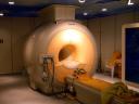
You have probably heard about CAT and MRI scans (produced thanks to machines like the one to the top right). So you know that these are techniques that doctors and scientists use to look inside the brain.
You have probably also heard about brain fitness and how important it is to keep a healthy brain to be protected against age-related and disease-related brain damages.
The question we ask here is the following: Can we use brain scans to evaluate how fit the brain is? Before we try to answer this question let’s start with the basics and try to understand how brain scans work.
Brain imaging, also called neuroimaging, allows one to produce images of the brain. There are different types of brain imaging: structural and functional. Structural imaging provides information about the shape and volume of the brain (CAT and MRI scans). Functional imaging shows the brain cells that are active when one performs a specific task (fMRI and PET scans).
 CT or CAT scans. Computed tomography or computed axial tomography is a technique that takes a large number of two-dimensional X‑rays images. These images are used to digitally compute 3D images of the inside of the brain. The most frequent reason for a head CT is to diagnose cerebrovascular accidents and intracranial hemorrhage. It is also often use to evaluate facial and skull fractures.
 MRI scans. MRI stands for Magnetic Resonance Imaging. This technique uses magnetic fields and radio waves to generate 2- or 3‑D images of the brain. MRI is used to detect tumors and other pathologies that affect the tissues of the brain (e.g., multiple sclerosis). Compared to CT, it is more precise and harmless to the patient (no potentially dangerous X‑rays). However, CT is much cheaper and more widely used.
 PET scans. PET stands for Positron Emission Tomography. It measures the emission generated by a short-lived radioactive tracer injected to the patient (using the bloodstream). The 2- or 3‑D images produced show brain activity. PET is used to detect tumors and for the diagnosis of brain diseases. Since the 1990s, fMRI has supplanted PET due to its low invasiveness, lack of radiation exposure, and relatively wide availability.
 fMRI scans. Functional Magnetic Resonance Imaging relies on the magnetic properties of oxygenated and deoxygenated hemoglobin. This technique produces images of changing blood flow in the brain associated with neural activity. The images show the brain structures activated during performance of different tasks. fMRI is used to detect early changes in the brain following strokes or other brain diseases.
What is brain reserve?
Now you know about imaging techniques. Our goal was to see whether we could use these techniques to evaluate whether a brain is fit or not. Fit brains are brains that have what scientists call cognitive brain reserve. It is more or less the capacity of the brain to resist the expression of symptoms in the face of existing neuropathology. In other words, people with more cognitive brain reserve can tolerate more pathologic changes before they show any symptoms. If you have healthy brain, that is if you have cognitive reserve, you will still get older and you may still get Alzheimer disease. But brain reserve will help delaying the effects of age and the onset of dementia. You may be wondering: “What can I do to get some cognitive reserve? Here are 2 factors that seem crucial:
- Education (see Snowdon et al., 1989 or Katzman, 1993)
- Level of intellectual stimulation (through your job or your leisure activities) (see Scarmeas et al., 2001; Verghese et al., 2003)
It looks like education or intellectual stimulation may increase brain reserve by increasing the density of the connections between brain cells (that is by increasing synapses between neurons). For more info about cognitive reserve see Alvaro interview with researcher Dr. Yaakov Stern.
Can you image brain reserve in the brain?
In a recent study, Perneczky and colleagues (2006) used PET to explore the effect of cognitive reserve on Alzheimer’s disease. They scanned the brain of 93 patients with mild Alzheimer’s disease and 16 healthy controls.
It was expected that people with the more severe Alzheimer’s disease pathology would show less cerebral blood flow, that is less activity, in the regions affected by the disease. Remember that blood flow in the brain is what PET measures.
Perneczky hypothesized that patients with more years of schooling would have more pronounced deficits in regions typically affected by the pathology of Alzheimer’s disease.
How could this be? Didn’t I tell you earlier that education contributes to brain reserve?
Here is the reasoning: say that Ms A. has a low level of brain reserve. She has developed Alzheimer’s 2 years ago. She doesn’t have much pathology in her brain yet and her symptoms (memory problems, etc.) correspond to a mild stage of Alzheimer’s disease. Ms B. has a high level of brain reserve. She has developed Alzheimer’s 5 years ago. She has a high level of pathology in her brain. However, thanks to her brain reserve, she only shows symptoms corresponding to a mild stage of Alzheimer’s disease.
Results of this PET study show that indeed patients with more education (such as Ms B.) consistently had more pronounced deficits in regions typically affected by the pathology of Alzheimer’s disease compared to patients with less years of schooling (such as Ms A.)
These findings suggest that education is associated with brain reserve and that people with higher education can cope with brain damage for a longer time.
What if one doesn’t have higher education…Is it too late to build cognitive brain reserve? The good news is that it is NOT too late! Education is not the only factor. One can always find ways to get enough mental stimulation by choosing our jobs and engaging in leisure activities such as reading, learning new things, going to museums, etc.
 — This article was written by Pascale Michelon, Ph. D., for SharpBrains.com. Dr. Michelon has a Ph.D. in Cognitive Psychology and has worked as a Research Scientist at Washington University in Saint Louis, in the Psychology Department. She conducted several research projects to understand how the brain makes use of visual information and memorizes facts. She is now an Adjunct Faculty at Washington University, and teaches Memory Workshops in numerous retirement communities in the St Louis area.
Share this:

5 Comments
About SharpBrains
Top Articles on Brain Health and Neuroplasticity
Top 10 Brain Teasers and Illusions
Got the book?

Really looking inside the brain?
The studies cited all use proxies for cognitive reserve (e.g. years of education), but these indirect measures crudely approximate the brain effects of such lifetime experiences. We really need neuroimaging techniques capable of directly measuring the expression of neural networks associated with cognitive reserve. We would then be in a position to prospectively test the effects of activities purported to increase reserve.
Hi Joshua,
I agree with your comment. Neurogenesis and the like is really hard to measure in humans. Small and colleagues (2007) used MRI to try to estimate the effect of physical activity on neurons formation (which contributes to cognitive reserve). However, they again used a proxy as they could not directly count the new neurons…
Hi, Your article was included in the First Carnival of Tech at http://www.technologymatter.com/2008/02/first-carnival-of-tech.html.
Thanks!
Neuroimaging is such an essential part of modern medicine today, your website is bringing much needed attention to this area.
Thank you, Chris!