A new era of brain cartography, powered by neuroimaging and machine learning
– M GLASSER, D VAN ESSEN/WASHINGTON UNIVERSITY
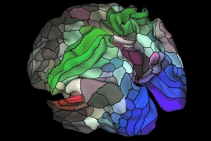
Human brain mapped in unprecedented detail (Nature):
“Think of a spinning globe and the patchwork of countries it depicts: such maps help us to understand where we are, and that nations differ from one another. Now, neuroscientists have charted an equivalent map of the brain’s outermost layer — the cerebral cortex — subdividing each hemisphere’s mountain- and valley-like folds into 180 separate parcels.
Ninety-seven of these areas have never previously been described, despite showing clear differences in structure, function and connectivity from their neighbours. The new brain map is published today in Nature…
“While the focus of this work was on creating a beautiful, reliable, average brain template, it really opens up the possibility to further explore the unique intersection of individual talents with intellectual and creative abilities — the things that make us uniquely human,” says Rex Jung, a neuropsychologist at the University of New Mexico in Albuquerque.
Study: A multi-modal parcellation of human cerebral cortex (Nature; opens PDF)
- Abstract: Understanding the amazingly complex human cerebral cortex requires a map (or parcellation) of its major subdivisions, known as cortical areas. Making an accurate areal map has been a century-old objective in neuroscience. Using multi-modal magnetic resonance images from the Human Connectome Project (HCP) and an objective semi-automated neuroanatomical approach, we delineated 180 areas per hemisphere bounded by sharp changes in cortical architecture, function, connectivity, and/or topography in a precisely aligned group average of 210 healthy young adults. We characterized 97 new areas and 83 areas previously reported using post-mortem microscopy or other specialized study-specific approaches. To enable automated delineation and identification of these areas in new HCP subjects and in future studies, we trained a machine-learning classifier to recognize the multi-modal ‘fingerprint’ of each cortical area. This classifier detected the presence of 96.6% of the cortical areas in new subjects, replicated the group parcellation, and could correctly locate areas in individuals with atypical parcellations. The freely available parcellation and classifier will enable substantially improved neuroanatomical precision for studies of the structural and functional organization of human cerebral cortex and its variation across individuals and in development, aging, and disease.
To learn more:
Share this:

About SharpBrains
Top Articles on Brain Health and Neuroplasticity
Top 10 Brain Teasers and Illusions
Got the book?